Even the mucous membranes of the larynx and in particular of the vocal cords can degenerate form of malignant tumors (predominantly squamous cell carcinomas).
This phenomenon occurs mostly in patients who smoke or who drink alcohol are chronically.
The degenerative process is a long process, characterized by several stages that are collected in the generic definition of Squamous intraepithelial lesions (Sils). Some types of SILs are self-limiting and reversible, Some are stable, other proceeding inexorably towards the SCC, Despite an adequate follow-up and treatment.
The most frequently used classification is the one offered by the World Health Organization (Who) in 2005, distinguishing laryngeal precancerous in:
1-Squamous cell hyperplasia: We observe an increased cell proliferation that can affect the level cell carcinoma of the skin, in this case we speak of Acanthosis, or prebasali or basal layers; the architecture is smooth and lacks atypia
2-Mild dysplasia: architectural alteration accompanied by atypia, limited to the lower third of the epithelium
3-Moderate dysplasia: interesting architectural alteration the middle third of the epithelium, with prominent nucleoli and nuclear abnormalities presenting cells, no abnormal mitosis. The lesions may be associated with Actinic
4-Severe dysplasia: interesting more than two-thirds of the epithelium, with prominent architectural anomalies, atypia, obvious nuclear abnormalities, loss of maturation, nuclear pleomorphism, bizarre nuclei. We observe an increased number of mitosis. Severe dysplasia has the same risk of developing invasive carcinoma of carcinoma in situ
5-Carcinoma in situ: that is a malignant transformation that does not invade the basement membrane
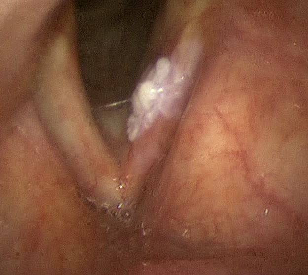
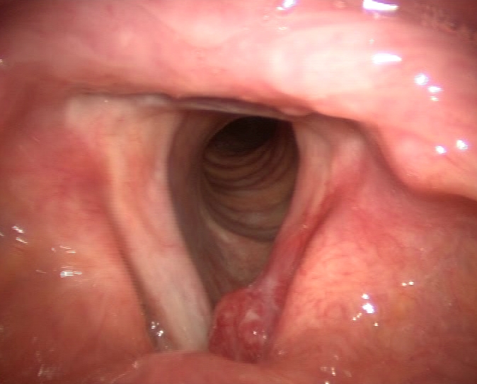
The precancerous and malignant tumors of the vocal cords can cause interference to the voice now (dysphonia) and so often come to the attention of the doctor still in early stage.
All ’ fibroscopico examination appear as a small whitish aerial detected (Leukoplakia) or reddish (erythroplakia) on the surface of the vocal cord.
Every patient who suffers from dysphonia for over 2 weeks should be evaluated by an Otolaryngologist with exam fibroscopico.
An expert clinical eye and further investigations (autofluorescence, NBI, TC, RMN) can help distinguish a frankly malignant precancerous lesion.
In the case of small precancerous lesions that have features of malignancy can implement a meticulous "attitude with periodic revaluations fibroscopiche condition only on condition that the patient WILL ABOLISH smoking cigarettes and drinking alcohol ’. In some cases you may see a lesion regression and avoid surgery.
In case the lesion appears suspect or the patient is not cooperating is useful to remove the lesion using a direct intervention in microlaringoscopia in suspension (MLSD), with help of CO2 LASER, to be carried out under general anesthesia. Even if early intervention l ’ malignant tumors may be curative and may not require further treatment but only careful periodic revaluations.


")