Nasal Endoscopy is a non-invasive outpatient procedure of exploration of the nasal cavity by means of a rigid endoscope or fiberscope. The procedure is generally well tolerated and causes a slight annoyance. Sometimes you may need to use local anesthetic and/or decongestant to reduce swelling of the mucous membranes and make more accessible the exploration of the nasal cavities. Continue reading
ENT Exams
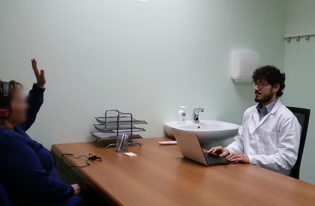
Head impulse test
The head impulse test is a clinical examination is very useful in the evaluation of patients with dizziness or balance deficit.
And’ a noninvasive test very quickly but not always easy to evaluate.
It asks the patient to fix a point in the vicinity of the eyes of the operator (the tip of the nose or a shirt button) and they make the patient's head of short, but very quickly to the right or left unpredictably.
Under normal conditions the brain uses information from the sight and the organ of balance and manages to keep the fixed pupils on target.
If the organ of balance does not work correctly, Turning her head to the side sick, the pupil will fail to stay on target and will tend to go sideways; later the brain realizes the mistake and fixes his gaze with a quick movement of the pupil (saccade).
The impedance
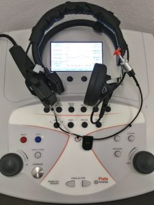 The impedance is a quick review, painless and non-invasive way that easily allows to objectify the presence of fluid in the middle ear (payment endotimpanico), changes in air pressure in the middle ear and to assess the presence of stapedial reflexes. Continue reading
Head shaking test
Movie showing the head shaking test positive, triggering important swing-right nystagmus
L’esame otovestibolare
Dizzy and the Unsteadiness ’ are a very frequent problem, can affect both young and old, both healthy people who with Comorbidities and can be extremely disabling.
Our balance depends on a great number of factors, in fact, our brain processes much information coming from 2 organs of ’ balance (placed in the ’ inner ear), from view, from our touch and our joints. Integrating this information we are able to perceive our position in space. When one of the mechanisms at play or the brain's ability to integrate information are compromised here is that you can check the ’ instability and dizziness.
The shapes that depend on ’ organ of ’ balance ’ are framed and followed by Otolaryngologist, While those who depend on our brains are followed by neurologist. Only rarely do the shapes are quite separate so the patient often giddy is evaluated for the first time by ’ Otolaryngologist also for neurological forms.
L ’ Otolaryngologist has thereby the responsibility first of all to identify the shapes which can pose a real risk to the life of the patient (some forms of stroke may present with Vertigo) and then differentiate into shapes of Central and peripheral set a correct diagnosis. Continue reading
L ’ tone audiometric test
 L ’ tonal audiometric allows decrease of hearing by quantifying ’ l ’ objectify extent of hearing loss and which frequencies being affected. And’ can also study separately the functionality of the auditory nerve and sound transmission system (outer ear, tympanic membrane, auditory ossicles and the middle ear).
And’ a simple exam ’, non-invasive and short-lived.
The patient is placed in a quiet or soundproofed and are placed headphones through which are heard of pure tones (sounds with a specific frequency) a ’ intensity note. The patient is asked to report when it is able to hear sound. Is then marked on a path the minimum intensity at which the subject is able to hear the sounds, distinguishing the individual frequencies and two ears.
Videonistagmografia
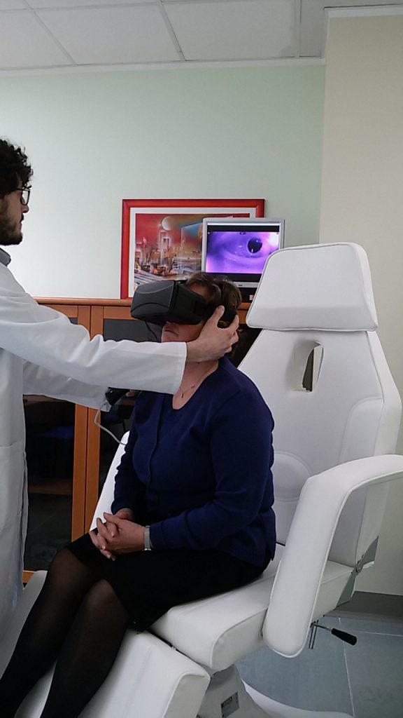 In healthy person, l ’ ’ organ of balance (vestibular system) It also plays a key role in ’ assist the brain in controlling eye movements so as not to lose the setting of an object despite abrupt head movements.
In pathology the vestibular system can cause abnormal eye movements called nystagmus.
L ’ careful evaluation of these movements is spontaneous that caused is essential to frame the dizziness and ’ instability. Being eye-movements controlled by the brain from the vestibular system it is important to try to exclude as much as possible, l ’ central task: It then tries to put the patient in a position not to see the ’ environment. To achieve this condition you can use goggles Frenzel with strongly miopizzanti that waste the patient lenses every visual reference.
L ’ technological evolution is represented by videonistagmografia: a mask with camera with Infrared Illuminator. Once worn the patient will be in complete darkness while l ’ examiner will investigate and possibly record the movements of eyes evaluating in a monitor.
Registering with videonistagmografia Rotary nystagmus, physiologic in healthy person Continue reading
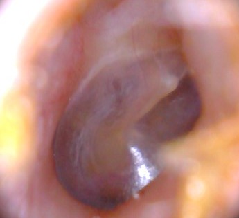
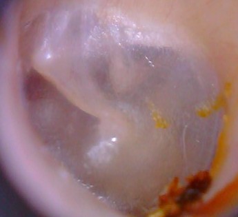
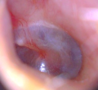
Otoscopy
L ’ Otoscope is an instrument with a graft for disposable ear cones, a light and a magnifying glass.
Allows you to observe the external ear canal and tympanic membrane in a much smoother. And’ simply effectual a slight pull towards the external ’ and later on the ear and place the instrument.
And’ possible to exclude the presence of earwax plugs, of narrowing of the external ear canal (Exostoses and osteomas), exclude external otitis, evaluate the tympanic membrane integrity ’, exclude miringiti, acute otitis media and effusive.
The exam can be done easily in children.
Fiberoptic in evaluating dysphagia
Dysphagia, that is the difficulty in swallowing foods, can have many causes and may be caused by an obstacle or laryngopharyngeal level or oesophageal.
L ’ Otolaryngologist plays an important role in the evaluation of dysphagic patients, both in identifying the cause ’ that in ’ food standards setting to minimize risks due to dysphagia. In fact dysphagic patients have a higher risk of inhalation of foods: If swallowing is not effective foods may accidentally penetrate the Airways. L ’ inaction can cause serious lung infections.
The standard fiberoptic allows to evaluate l ’ Anatomy and the correct capacity of the larynx to prevent food from entering the trachea.
Also you can see the pharynx and larynx in real time while the patient ingests liquid foods and semi-solid colored. In this way you can have a clear idea of the level of dysphagia and introduce measures to minimise the risk of pneumonia.
Fiberoptic in the assessment of snoring
All patients with snoring and obstructive sleep apnea syndrome (OSAS) should conduct an assessment ORL with fiberoptic.
In patients with snoring is initially made a standard fiberoptic in order to exclude anatomical issues that might affect or cause the pathology (nasal septum deviation, hypertrophy of lingual base, Palatal PTOSIS)
Then it is important to perform the maneuver of Muller: It requires the patient to a ’ forced inspiration keeping my mouth shut and nostrils plugged. The negative pressure that is created in the pharynx simulates pharyngeal collapse that occurs in sleep with reduced muscle tone.
This procedure allows to simulate, non-invasively, the collapse of the Airways that occurs during sleep.
Identify the anatomical problem that can affect your snoring can be set with the patient proper treatment plan


")