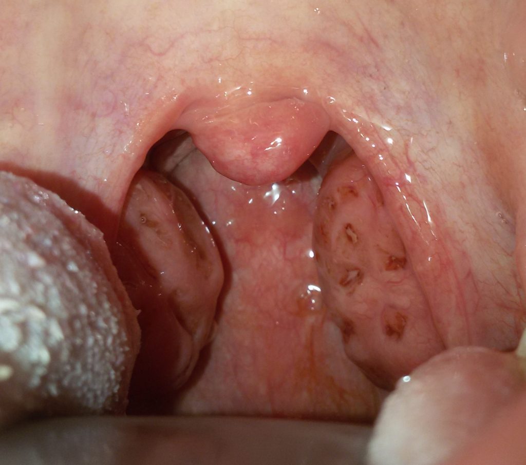
Tonsillectomy is surgery to remove ’ of the palatine tonsils. And’ a routine procedure, safe, that is currently being carried out under general anesthesia.
The tonsils are removed or infectious diseases (Recurrent acute tonsillitis) or for airway obstruction problems or suspected malignancy.
The criteria that lead to a correct indication for tonsillectomy differ substantially between children and adults.
 Rhinoplasty is the surgery that allows ’ aesthetic nose correction.
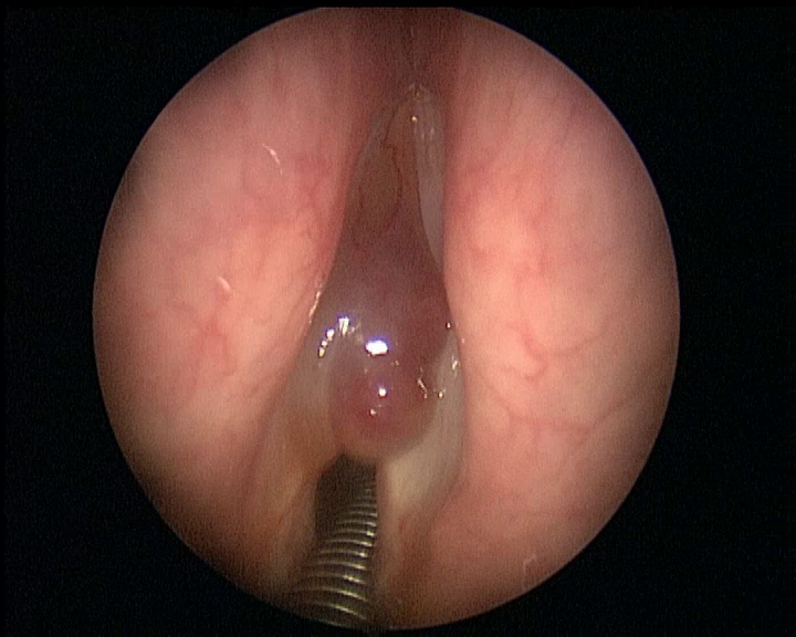
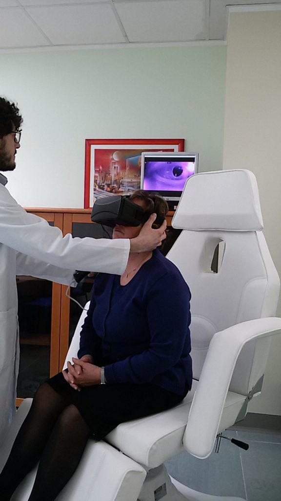 In healthy person, l ’ ’ organ of balance (vestibular system) It also plays a key role in ’ assist the brain in controlling eye movements so as not to lose the setting of an object despite abrupt head movements.
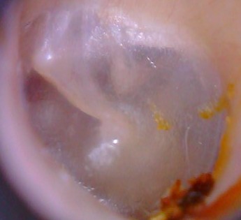
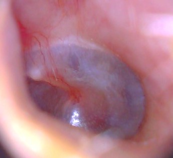
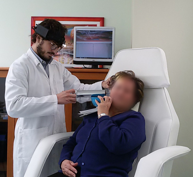 L ’ outer ear physiologically produces ceruminose secretions that protect it from infection and microtrauma. Earwax is spontaneously spontaneously delivered from the skin to the outer ’ where will later be removed with normal hygiene. Earwax mingling to de-epithelialised skin can build up causing the dreaded Earwax plugs. Some people are very prone to the formation of earwax plugs, are known risk factors: l ’ hypersecretion of earwax, l ’ use of cotton buds (crushing Cerumen toward the eardrum), l ’ routine use of earphones, diseases that cause increased skin de ’ (keratitis, Psoriasis), narrow ear canal or shrink from osteomas and Exostosis. Also with age ear wax tends to be harder and so it is more prone to the formation of caps with ’ advancing years. Ear cerumen has a particular feature: is hygroscopic, namely swells when it's wet with water. And’ frequent that patients report feeling much less, suddenly, After taking a shower. Earwax plugs can give various symptoms: conductive hearing loss, autophony (hear their voices reverberate), Tinnitus, pain (ear pain) If they appear over infections. A simple otoscopy enables you to diagnose Cap. There are different techniques to remove plug of earwax. The simplest and the least disturbing to the patient is to resort to washing earphones: a jet of water ’ (at body temperature) sprayed in the external ear canal insinuates itself between the skin and Earwax him taking it off from. In the event that you suspect a ’ infection (already have pain) or there is a known perforation of tympanic basolateral or any other condition of ’ ears,or washing is however not recommended. You can remove the Earwax with hooks, Anse, spatulas, Hartmann's pliers or with suction under careful Visual inspection.
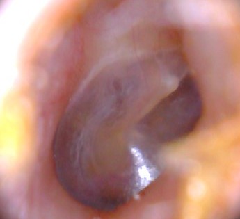


")